
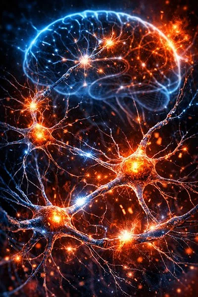
Photobiomodulation Benefits for Parkinson’s Disease
Is It Time to Consider Photobiomodulation As a Drug Equivalent?
by Tiina Karu, PhD, DrSci
2013
Vital Forces Summary (Study below)
In this article, Tiina Karu pulls together a wide range of studies that explicitly identifies Parkinson’s disease as one of the most promising clinical applications for photobiomodulation. Photobiomodulation is revealed as a potential disease-modifying therapy rather than just a symptomatic treatment. Karu shows evidence that:
PBM prevents mitochondrial dysfunction
PBM prevents dopamine loss
PBM restores axonal transport
PBM stimulates neuronal respiration
In a 2010 study, Shaw documents specific benefits of red and near-infrared (NIR) light:
Prevention of mitochondrial dysfunction
Protection of midbrain dopaminergic neurons
Reduction of dopamine loss
These benefits demonstrate true neuroprotection, not merely symptomatic improvement, and that Photobiomodulation targets the core pathology of Parkinson’s disease: mitochondrial failure and dopaminergic neuron degeneration.
Two 2009 articles by Trimmer, Bennett and others refer to Trimmer’s study with Red and NIR light on cybrid neurons in Parkinson’s Disease. They document the following benefits:
Restored axonal transport
Normalized mitochondrial movement
Stimulated cellular respiration
Reversed cellular degeneration associated with Parkinson’s pathology
These studies identified reduced axonal transport as a major degenerative mechanism in Parkinson’s disease. They also establish a cellular model demonstrating mitochondrial impairment relevant to sporadic Parkinson’s disease. They show that PBM can reverse intracellular transport failure, a critical contributor to neuronal degeneration.
When all the studies and articles from Karu’s article are taken together, we can see the scope of benefits to Parkinson’s Disease:
Mitochondrial Effects
Prevention of mitochondrial dysfunction
Increased mitochondrial respiration
Improved mitochondrial mobility within neurons
Neuronal Preservation
Protection of dopamine-producing neurons
Reduced dopamine depletion
Preservation of neuronal integrity
Axonal Transport
Restoration of axonal transport mechanisms
Reversal of transport-related neurodegeneration
Disease-Modifying Potential
Acts on upstream degenerative mechanisms, not downstream symptoms
Preferential effectiveness in stressed or diseased cells
Suggests neuroprotective and neurorestorative, not merely palliative, action
Is It Time to Consider Photobiomodulation As a Drug Equivalent?
by Tiina Karu, PhD, DrSci
The question of whether photobiomodulation should be used as a drug equivalent arose in my mind after listening to presentations at the recent conference of the World Association for Laser Therapy (WALT)-2012 (Gold Cost City, Australia), and later at home when searching MEDLINE® for the years 2009–2012. Photobiomodulation (earlier terms: low level laser therapy, LLLT, laser biostimulation) has been used in clinical practice for > 40 years by now, and its action mechanisms on cellular and molecular levels have been studied for > 30 years. Enthusiastic medical specialists successfully used photobiomodulation in treating healing-resistant wounds and ulcers (e.g., chronic diabetic ulcers), in pain management, and in spinal cord and nervous system injuries when other methods had had limited success.1 However, photobiomodulation is still not a part of mainstream medicine. The goal of the present Editorial is to highlight some important recent developments in clinical applications and in studies of cellular and molecular mechanisms behind the clinical findings.
One of the impressive and perspective challenges for photobiomodulation is its use in cases of Parkinson’s disease. Research in recent years evidenced that neuroprotective treatment with red and near infrared radiation (NIR) prevented mitochondrial dysfunction and dopamine loss in Parkinson’s disease patients.2 In another set of experiments, NIR normalized mitochondrial movement and axon transport, as well as stimulating respiration in cytoplasmic hybrid (‘‘cybrid’’) neurons.3,4 It is important to recall that reduced axonal transport contributes substantially to the degeneration of neuronal processes in Parkinson’s disease.
Another development in recent years is the successful stimulation of stem cells with red and NIR radiation. One example is the treatment of myocardial infarction. The heart has been considered a post-mitotic organ lacking the capacity for self-renewal after injury. Surprisingly enough, human cardiac stem cells, in combination with bone marrow mesenchymal stem cells, were found to reduce infarct size and restore cardiac functions after myocardial infarction.5 This positive effect can even be increased by irradiation of stem cells. Mesenchymal stem cells were derived from bone marrow and adipose tissue, and stimulated by irradiation at k = 810 nm. Implantation of irradiated cells into the infarcted rat heart resulted in an *50% decrease in cardiac infarct size.6 An increase in proliferation rates and membrane potential was established after 532 nm irradiation of adipose tissue-derived stem cells.7 A recent review8 summarized data about enhancement of the proliferation of various cultured cell lines, including stem cells, as well as cell lines used for the production of viral vaccines and hybrid cell lines. The review8 underlined that photobiomodulation improves the proliferation of cells without causing any cytotoxic effects. One has to emphasize that laser therapy shares none of the risks associated with stem cell therapy, requires no anesthesia, and is painless.9 The optimal light parameters in this review8 were found to be as follows: doses were 0.5–4.0 J/ cm2 and wavelengths ranged from 600 to 700 nm. It is important to recall that, in this particular wavelength range, two peaks in absorption and action spectra connected with activation of cytochrome c oxidase (the primary photoacceptor for photobiomodulation effects) are situated.10 The peak at 620 nm belongs to reduced CuA, and that at 680 nm, to oxidized CuB atoms in cytochrome c oxidase molecule.11
The treatment of vitiligo (a depigmentary disorder) remains a challenge for clinical dermatologists. He-Ne laser irradiation was found to stimulate melanocyte proliferation.12 The expression of phosphorylated cyclic-adenosine monophosphate (AMP) response element-binding protein, an important regulator of melanocyte growth, was upregulated by He-Ne laser treatment. He-Ne laser irradiation imparted a growth stimulatory effect on functional melanocytes via mitochondria-related pathways.12
Irradiation with red light caused gene and noncoding RNA regulation for photoacceptor protection in the retina. This finding may open a new challenge for photobiomodulation.13
One of the major dose-limiting effects of chemotherapy drugs is oral mucositis of treated patients. Oral mucositis can affect up to 100% of patients undergoing high-dose chemotherapy and hematopoietic stem cell transplantation. Photobiomodulation can improve tissue repair and immune response in these patients.14
Photobiomodulation has been shown to improve functional outcome after surgical intervention to repair injured nerves. LED irradiation at 810 nm accelerated functional recovery and improved the quality of nerve regeneration after autograft repair of severely injured peripheral nerves.15
Forehead treatments with NIR reversed major depression and anxiety.16 Transcranial NIR laser therapy was investigated as a new neuroprotective treatment for acute ischemic stroke.17 The authors of these studies believe that the irradiation promoted functional and behavioral recovery via cellular mitochondrial mechanisms, as well as by enhancing cerebral blood flow.
Photobiomodulation altered cardiac cytokine expression following acute myocardial infarction,18 as well as protected cardiomyocytes from hypoxia and reoxygenation injury via nitric oxide-dependent mitochondrial mechanisms.19
The analgetic effects of photobiomodulation have been studied for years, and are documented rather well.20 Light treatment (k = 635 nm) of open skin wounds of corticosteroidtreated diabetic rats was useless, as compared with nonsteroid laser treatment, in which case a significant acceleration of epitalization and collagen synthesis was observed.21 This finding could probably explain why in some clinical cases the laser treatment of wounds has a low efficiency. At the same time, irradiation at 660 nm was effective for collagen production in diabetic wounded fibroblasts.22
I will conclude by discussing ‘‘mitochondrial mechanisms of photobiomodulation,’’23 the term used widely in recent years for molecular and cellular mechanisms of light action. The rather old suggestion10,11 that the photoacceptor for photobiomodulation effects is cytochrome c oxidase has been confirmed by now.24 The new data25 support the old conclusion that photobiomodulation is more pronounced in ill or otherwise stressed cells, as compared with healthy cells with plenty of oxygen available.26 The terminal enzyme of the mitochondrial respiratory chain and its electronic excitation by light with proper parameters causes retrograde light-sensitive cellular signaling events to transport the light signal from mitochondria to the nucleus to cause gene expression.27 The gene expression events caused by irradiation are confirmed, and have been studied in more detail in recent years.28,29
Acknowledgments
This study was supported by the grant No 11-02-00295 RFFI.
Institute of Laser and Information Technologies of Russian Academy of Sciences, Moscow, Russian Federation.
References
1. Tuner, J., and Hode, L. (2011). The laser therapy handbook. Gra¨ ngesberg: Prima Books.
2. Shaw, V.E., Spana, S., Ashkan, K. Benabid AL, Stone J, Baker GE, and Mitrofanis, J. (2010). Neuroprotection in midbrain dopaminergic cells in MPTP-treated mice after near-infrared light treatment. J. Comp. Neurol. 518, 25–40.
3. Trimmer, P.A., and Benett, J.P. (2009). The cybrid model of sporadic Parkinson’s desease. Exp. Neurol. 218, 320–325.
4. Trimmer, P.A., Schwarz, K.M., Borland, M.K., De Taboada, L., Streeter, J., and Oron, U. (2009). Reduced axonal transport in Parkinson’s disease cybrid neurites is restored by light therapy. Mol. Neurodegener. 4, 26–40.
5. Williams, A.R., Hatzistergos, K.E., Addicott, B., McCall, F., Carvalho, D., Suncion, V. and Morales, A.R., Da Silva, J., Sussman, M.A., Heldman, A.W., and Hare, J.M. (2013). Enhanced effect of combining human cardiac stem cells in bone marrow mesenchymal stem cells to reduce infarct size and to restore cardiac function after myocardial infarction. Circulation 127, 213–223.
6. Tuby, H., Maltz, L., and Oron, U. (2008). Implatation of low-level laser irradiated mesenhymal stem cells into the infarcted rat heart is associated with reduction in infarct size and enhanced angiogenesis. Photomed. Laser Surg. 27, 227–234.
7. Anwer, A.G., Gosnell, M.E., Perinchery, S.M., Inglis, D.W., and Goldys, E.M. (2012). Visible 532 nm laser irradiation of human adipose tissue-derived stem cells: effect on proliferation rates, mitochondria membrane potential and auto- fluorescence. Lasers Surg. Med. 44, 769–778.
8. AlGhandi, A., Kumar, A., and Moussa, N.A. (2012). Low- level laser therapy: a useful technique for enhancing the proliferation of various cultured cells. Lasers Med. Sci. 27, 237–249.
9. Malliaras, K., Zhang, Y., Senfeld, J., Galang, G., Tseliou, E., Cheng, K., Sun, B., Aminzadeh, M., and Marban, E. (2013). Cardiomyocite proliferation and progenitor cell recruitment underlie therapeutic regeneration after myocardial infraction in the adult mouse heart. EMBO Molec. Med. 5, 191–209.
10. Karu, T.I., Pyatibrat, L.V., Kolyakov, S.F., and Afanasyeva,
N.I. (2005). Absorption measurements of a cell monolayer relevant to phototherapy: reduction of cytochrome c oxidase under near IR radiation. J. Photochem. Photobiol. B Biol., 81, 98–106.
11. Karu, T.I. (2010). Multiple roles of cytochrome c oxidase in mammalian cells under action of red and IR-A radiation. IUBMB Life 62, 607–610.
12. Lan, C.C.E., Wu, C.-S., Chiou, Y.-H., Chiang, T.Y., and Yu,
H.S. (2009). Low-energy He-Ne laser induced melanocyte proliferation via interaction with type IV collagen: visible light as a therapeutic option for vitiligo. Br. J. Dermatol. 161, 273–280.
13. Natoli, R., Zhu, Y., Valter, K., Bisti, S., Eells, J., and Stone, J. (2010). Gene and noncoding RNA regulation underlying photoreceptor protection: Microarray study of dietary antioxidant saffron and photobiomodulation in rat retina. Mol. Vis. 16, 1801–1822.
14. Bjordal, J.M., Bensadoun, R.-J., Tuner, J., Frigo, L., Gjerde, K., and Lopes-Martins, R.A. (2011). A systematic review with meta-analysis of the effect of low-level laser therapy (LLLT) in cancer therapy-induced oral mucositis. Support. Care Cancer 19, 1069–1077.
15. Moges, H., Wu, X., McCoy, J., Vasconcelos, O.M., Bryant, H., Grunberg, N.E., and Anders, J.J. (2011). Effect of 810 nm light on nerve regeneration after autograft repair of severely injured rat median nerve. Lasers Surg. Med. 43, 901–906.
16. Schiffer, F., Johnston, A.L., Ravichandean, C., Polcari, A., Teicher, M.H., Webb, R.H., and Hamblin, M.R. (2009). Psychological benefits 2 and 4 weeks after a single treatment with NIR to the forehead: a pilot study of 10 patents with major depression and anxiety. Behav. Brain Funct. 5, 46.
17. Lapchak, P.A. (2010). Taking a light approach to treating acute ishemic stroke patients: transcranal near infrared laser therapy in translational science. Ann. Med. 42, 576–586.
18. Yang, Z., Wu, Y., Zhang, H., Jin, P., Wang, W., Hou, J., Wei, Y., and Hu, S. (2011). Low–level laser irradiation alters cardial cytokine expression following acute myocardial infraction: a potential mechanism for laser therapy. Photomed. Laser Surg. 29, 391–398.
19. Zhang, R., Yasushi, M., Pratt, P.F., Lohr, N., Warltier, D.C.,
Whelan, H.T., Zhu, D., Jacobs, E.R., Medhora, M., and Bienengraber, M. (2009). Near infrared light protects cardiomyocytes from hypoxia and reoxygenation injury by a nitric oxide dependent mechanism. J. Mol. Cell Cardiol. 46, 4–14.
20. Chow, R., Arwati, P., Laakso, L., Bjordal, J., and Baxter, G. (2011). Inhibition and releance to analgetic affects: A systematic review. Photomed. Laser Surg. 29, 365–381.
21. Gal, P., Mokry, M., Vidinsky, B., Kilik, R., Depta, F., Harakalova, M., Longauer, F., Mozel, S., and Sabo, J. (2009). Effect of equal daily doses achieved by different power densities of low-level laser therapy at 635 nm on open skin wound healing in normal and corticosteroid-treated rats. Lasers Med. Sci., 24, 539–547.
22. Ayuk, S.M., Houreld, N.N., and Abrahamse, H. (2012). Collagen production in diabetic wounded fibroblasts in response to low-intensity laser radiation at 660 nm. Diabetes Technol. Ther. 14, 1110–1117.
23. Karu, T.I. (2010). Mitochondrial mechanisms of photobiomodulation in context of new data about multiple roles of ATP. Photomed. Laser Surg., 28, 159–160.
24. Hamblin, M.R. (2010). Introduction to experimental and clinical studies using low level laser (light) therapy (LLLT). Lasers Surg. Med. 42, 447–449.
25. Masha, R.T., Houreld, N., and Abrahamse, H. (2012). Low- intensity laser irradiation at 660 nm and stimulates cytochrome c oxidase in stressed fibroblast cells. Lasers Surg. Med. 44, 429–434.
26. Karu, T., (2007). Ten lectures on basic science of laser phototherapy. Gra¨ ngesberg: Prima Books AB.
27. Karu, T., and Pyatibrat, L. (2011). Gene expression under laser and light-emitting diodes radiation for modulation of cell adhesion: possible applications for biotechnology. IUBMB Life 63, 747–753.
28. McDaniel, D.H., Weiss, R.A., Geronemus, R.G., Mazur, C., Wilson, S., and Weiss, M.A. (2010) Varying ratios of wavelengths in dual wavelength LED photomodulation alters gene expression profiles in human skin fibroblasts. Laser Surg. Med. 42, 540–545.
29. Masha, R.T., Houreld, N.N., and Abrahamse, H. (2013). Low intensity laser irradiation at 660 nm stimulates transcription of genes involved in electron transport chain. Photomed. Laser Surg. 31, 47–53.
Photomedicine and Laser Surgery Volume 31, Number 5, 2013
ª Mary Ann Liebert, Inc. Pp. 189–191
DOI: 10.1089/pho.2013.3510